Arthroscopic Knee Surgery
KNEE PROBLEMS
SURGICAL PROCEDURES
What is Arthroscopy?
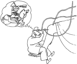 |
Illustration: Dusan Petricic
|
Arthroscopy is a surgical procedure which orthopaedic surgeons use to view, diagnose and treat problems inside the knee joint. The word arthroscopy comes from Greek words Arthros (joint) and Scopos (to look). The term literally means "to look inside the joint". In an arthroscopic operation, an orthopaedic surgeon makes a small incision in the patient's skin and then inserts the arthroscope, a miniature lens and lighting system, which magnifies and illuminates the structures inside the joint. This small instrument is approximately 5 mm in diameter. An intense, cool light is transmitted through fibreoptic cables to the end of the arthroscope that is inserted into the joint. By using a miniature colour video camera attached to the arthroscope, the surgeon is able to see the interior of the knee joint on a large television screen.
Why is Arthroscopy Necessary?
A knee joint is lined with the synovial lining, that normally produces minute amounts of joint lubricant and nutrient called synovial fluid, and contains dense and elastic tissue lining bone ends and the underside of the kneecap called articular cartilage, two tough cartilage cushions called medial and lateral menisci and fibre-like connecting tissue called ligaments. The articular cartilage, menisci and ligaments cushion the bones and stabilise the joint. Injuries and disease can damage synovial lining, bones, articular cartilage, menisci, ligaments, muscles and tendons.
 |
 |
|
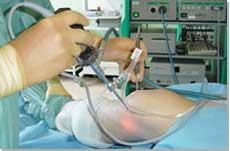
Arthroscopic knee surgery |
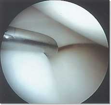
Normal meniscus and articulating surfaces |
Diagnosing knee joint injuries and other conditions begins with a thorough
medical history, physical examination, x-ray films (mainly for bone
pathology) and, quite often, MRI (magnetic resonance imaging, mainly
for soft tissue and articular cartilage injury). Further diagnosis using
arthroscopy may be required because it gives a precise, direct view
of the affected structures inside the joint. However, surgical intervention
for various problems affecting the inside of the knee joint is the main
reason for arthroscopy nowadays. Some of the most frequent conditions
found during arthroscopic examination of the knee joint are:
- Torn or degenerate meniscus or menisci (semilunar cartilage)
- Damaged joint surface (articular cartilage)
- Loose fragments of cartilage or bone (loose bodies)
- Maltracking and tilted patella (kneecap)
- Torn anterior cruciate ligament
- Inflammation of the joint lining (reactive synovitis)
How is Arthroscopy Performed?
Arthroscopic surgery requires the use of a hospital operating theatre. Before the procedure, a patient is given an anaesthetic. The tourniquet (tight cuff above the knee and around the thigh) may be used to temporarily interrupt the local circulation. The leg is painted, usually pink or light brown, to disinfect the skin surrounding the knee. Then a sterile fluid is introduced into the knee joint to expand it, making room for the arthroscope and surgical instruments. The surgeon makes a small incision in the skin and inserts the arthroscope. Usually 2 to 3 incisions, called portals, are necessary in order to examine the inside of the joint. A surgical instrument is used to probe various parts within the joint to determine the quality of the tissue and extent of the problem. If surgery is indicated, it is performed with specially designed manual or power-driven instruments that are inserted into the joint through the portals.
A selection of your arthroscopic images will be recorded and stored digitally, and kept as a part of your hospital record. We will give you a copy of the full arthroscopy report, with several representative intra-articular images. If you wish to have more detailed imaging information or the video recording please ask us to record your arthroscopic operation on a CD or bring your own USB stick.
We may ask you to sign an Imaging Consent Form (see below), according to the guide of the Department of Health, Nuffield Healths’ policy and the Data Protection Act 1998.
Complications
Although uncommon, complications do occur occasionally during or following diagnostic and surgical arthroscopy. They include excessive swelling or bleeding, skin and (extremely rarely) joint infection, phlebitis, blood clots (DVT) and very rarely technical problems with arthroscopic instruments. There are also anaesthetic risks, both during and after the procedure, which are minimal and infrequent.
When Can I Fly After Knee Arthroscopic Surgery?
There is no universal agreement as to when it is safe to travel by plane after knee arthroscopic surgery. It seems that most orthopaedic surgeons advise their patients not to fly for at least 2 weeks before and after straightforward arthroscopic surgery. Short flights do not seem to be a problem. However, long intercontinental flights are a potential problem as there is an increased incidence of spontaneous DVT (deep venous thrombosis), even in the young and healthy passengers. It is possible that sitting for long period of time, in a confined space and with very little leg room in economy class, could predispose to the development of deep venous blood clots, especially in people following recent knee surgery. The likelihood of developing postoperative leg blood clots depends on many different factors, including your general health, medical history, postoperative mobility and a number of risk factors (obesity, smoking, a history of DVT, etc.). If you have to travel by plane, before 2 weeks after your arthroscopy, it would be wise to contact your airline’s Medical Department and to ask them for advice. Also, please discuss this issue with your GP, as you may have to take prophylactic measures for several weeks.
Advice on travel-related DVT:
- Department of Health. Advice on travel-related DVT. DoH website, last modified on 12 March 2007.
- Mike J Clarke, et al. Compression stockings for preventing deep vein thrombosis in airline passengers. Cochrane Database of Systematic Reviews (Intervention Review), Issue 4, 2008 (re-published online with edits on 8 October 2008).
- People Logistics. When Can I fly? People Logistics Limited website. January 8, 2007
Downloads (general information):
- Chester Knee Clinic Arthroscopic Surgery Brochure

- Chester Knee Clinic Exercises After Arthroscopy
- Chester Knee Clinic Imaging Consent Form
Furtner information on arthroscopic surgery:
-
Nancy McKenzie, PhD. Knee Arthroscopic Surgery. The Encyclopedia of Surgery.
Pinterest CKC Arthroscopic Image Board
We are pleased to announce that we started posting a selection of original arthroscopic images from our daily clinical and surgical practice on Pinterest and Twitter. All images are anonymous and are intended to provide good quality visual information on typical knee problems but also normal knee anatomy, for educational purposes. We will aim to Tweet a few selected images each week but all images will be available on Pinterest Arthroscopy board. We may post some images on Instagram but we will see how things go with Pinterest. Please let us know your thoughts (office@kneeclinic.info)
Page last updated on 8 June 2014
Site last updated on: 28 March 2014
|
 Disclaimer: This website is a source of information
and education resource for health professionals and individuals
with knee problems. Neither Chester Knee Clinic nor Vladimir Bobic
make any warranties or guarantees that the information contained
herein is accurate or complete, and are not responsible for
any errors or omissions therein, or for the results obtained from
the use of such information. Users of this information are encouraged
to confirm the accuracy and applicability thereof with other sources.
Not all knee conditions and treatment modalities are described
on this website. The opinions and methods of diagnosis and treatment
change inevitably and rapidly as new information becomes available,
and therefore the information in this website does not necessarily
represent the most current thoughts or methods. The content of
this website is provided for information only and is not intended
to be used for diagnosis or treatment or as a substitute for consultation
with your own doctor or a specialist. Email
addresses supplied are provided for basic enquiries and should
not be used for urgent or emergency requests, treatment of any
knee injuries or conditions or to transmit confidential or medical
information. If you have sustained a knee injury or have a medical condition,
you should promptly seek appropriate medical advice from your local
doctor. Any opinions or information,
unless otherwise stated, are those of Vladimir Bobic, and in no
way claim to represent the views of any other medical professionals
or institutions, including Nuffield Health and Spire Hospitals. Chester
Knee Clinic will not be liable for any direct, indirect,
consequential, special, exemplary, or other damages, loss or injury
to persons which may occur by the user's reliance on any statements,
information or advice contained in this website. Chester Knee Clinic is
not responsible for the content of external websites.
Disclaimer: This website is a source of information
and education resource for health professionals and individuals
with knee problems. Neither Chester Knee Clinic nor Vladimir Bobic
make any warranties or guarantees that the information contained
herein is accurate or complete, and are not responsible for
any errors or omissions therein, or for the results obtained from
the use of such information. Users of this information are encouraged
to confirm the accuracy and applicability thereof with other sources.
Not all knee conditions and treatment modalities are described
on this website. The opinions and methods of diagnosis and treatment
change inevitably and rapidly as new information becomes available,
and therefore the information in this website does not necessarily
represent the most current thoughts or methods. The content of
this website is provided for information only and is not intended
to be used for diagnosis or treatment or as a substitute for consultation
with your own doctor or a specialist. Email
addresses supplied are provided for basic enquiries and should
not be used for urgent or emergency requests, treatment of any
knee injuries or conditions or to transmit confidential or medical
information. If you have sustained a knee injury or have a medical condition,
you should promptly seek appropriate medical advice from your local
doctor. Any opinions or information,
unless otherwise stated, are those of Vladimir Bobic, and in no
way claim to represent the views of any other medical professionals
or institutions, including Nuffield Health and Spire Hospitals. Chester
Knee Clinic will not be liable for any direct, indirect,
consequential, special, exemplary, or other damages, loss or injury
to persons which may occur by the user's reliance on any statements,
information or advice contained in this website. Chester Knee Clinic is
not responsible for the content of external websites.
[ back to top ]